
MammaFusion XR-BX
- contact:
- funding:
DFG, FWF
- Partner:
Prof. P. Baltzer, Medizinische Universität Wien, Österreich
- startdate:
2019
- enddate:
2022
To support the breast cancer diagnosis, suspicious structures that are only visible in the magnetic resonance tomogram of the breast are transferred to stereotactic X-ray mammograms. This should enable the more cost-effective and more widely available biopsy under X-ray control.
Introduction
The aim of this project is to establish a novel method for registration of magnetic resonance imaging (MRI) and stereotactic X-ray mammography based on an accurate simulation of the biomechanics of the female breast, which could reduce the cost-effectiveness of breast biopsy of MRI-detected breast lesions by up to 50%. We aim to transfer information from lesions that are only visible on MRI to mammographic target images with sufficient registration accuracy to perform mammogram-based lesion processing, thereby reducing the need for costly and time-consuming MRI biopsies.
Motivation
Breast MRI is currently the most sensitive method for detecting breast cancer. While there is no doubt that MRI detects cancer that has been overlooked by conventional methods such as mammography and ultrasound, additionally detected MRI lesions pose a workflow problem in the clinical management of breast disease. MRI follow-ups are costly and their interval is still controversial. MRI-guided procedures such as percutaneous biopsies or wire localization are even more expensive than MRI follow-ups and require special equipment and expertise. In addition, a survey by the European Society for Breast Imaging (EUSOBI) found a significant lack of availability of MRI interventions in Europe.
Consequently, new methods are required to translate MRI results into an efficient and practical clinical workflow: This approach should take into account both cost efficiency and limited accessibility.
In this project, a matching method between MRI and conventional X-ray mammograms and target images of X-ray biopsy will be developed and evaluated. The aim is to transfer MRI-based information to mammography projection images with sufficient registration accuracy to allow mammography-based lesion processing either by percutaneous biopsy or follow-up.
For this, the following scientific questions need to be answered:
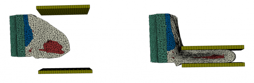
(1) The imaging conditions during stereotactic biopsy differ significantly from conventional mammography, as only target images are taken in the prone position. Since these views do not include the full breast shape, the challenge is to spatially align these views with full mammograms and/or MRI to provide diagnostic information for stereotactic biopsy.
(2) Identification of the necessary complexity of the biomechanical model that meets the clinical requirements in terms of both registration accuracy and computational time. For this purpose, it is important to investigate the correlation between registration accuracy, computation time and patient characteristics, since all three factors can significantly influence the robustness of the matching procedure.
(3) Identification of factors influencing the accuracy of agreement for stereotactic biopsy. These may consist of anatomical, clinical and biomechanical features that can be distinguished from those that only predict better or worse matching accuracy and those that can be incorporated into variations of the matching algorithm to improve its accuracy. This requires a sufficiently large clinical database. This will allow the identification of specific scenarios, including patient and lesion characteristics, associated with different matching outcomes that could be addressed by tailoring the complexity of the matching procedure to the specific case.
The IPE develops the registration method and carries out the evaluation of the method together with the University Hospital of Vienna. For this we use our developments in the field of model-based multimodal image registration, see Technologies.
Technology: Medical Image Processing